



Molecular residual disease (MRD) research

Explore NGS-based whole-genome sequencing and other methods for molecular residual disease research detection

What is molecular residual disease?

Molecular residual disease (MRD) refers to the presence of residual disease identified by tumor-derived molecular signals in solid tumor research. These signals may be present at levels below the detection limits of conventional disease assessment methods, including imaging. A related concept, minimal or measurable residual disease, has historically been used in hematologic malignancies to describe low levels of residual malignant cells detected using cell-based methods. While minimal and molecular residual disease research approaches are conceptually similar, molecular residual disease in solid-tumor research focuses on identifying tumor-derived molecular signals from circulating tumor DNA (ctDNA).

ctDNA represents a small fraction of the broader pool of cell-free DNA (cfDNA) found in blood samples. Using highly sensitive technologies such as polymerase chain reaction (PCR) and next-generation sequencing (NGS), researchers can detect and monitor these tumor-derived DNA fragments through a simple blood draw. Because cfDNA has a short biological half-life, NGS-based liquid biopsy testing can provide a real-time molecular snapshot of disease dynamics in research studies.1-3

How is molecular residual disease detected?

MRD can be detected in blood and other biological samples, depending on the research application and cancer type. MRD detection methods include PCR-based methods, targeted NGS, and whole-genome sequencing (WGS). The choice of detection method depends on the goals of the research application, assay design, and biomarkers being evaluated. Analytical sensitivity varies among MRD detection methods and depends on assay design and other analytical performance characteristics. These factors collectively influence a method’s ability to detect low levels of residual disease.2,4,5

Researcher in wet lab inspecting sample

Molecular residual disease detection method comparison

Summary table comparing representative reported limit of detection (LoD), molecular biomarkers measured, primary strengths, and limitations of MRD detection methods.2,5-11

Detection method Representative reported limit of detection (LoD)a Representative molecular biomarkers Primary strengths Primary limitations
Whole-genome sequencing (WGS) ~10−5 tumor fraction; ppm-level (~10−6) detection reported with newer tumor-informed WGS approaches Genome-wide tumor-derived variants, including single nucleotide variants (SNVs) and copy-number alterations (CNAs); signals can be aggregated across thousands of variants Genome-wide signal aggregation enables ultrasensitive detection at very low tumor fractions Higher sequencing and computational requirements and historically higher per-sample costs compared to methods such as targeted NGS and qPCR
Targeted next-generation sequencing (NGS) 10−4 to a few × 10−6 reported as ctDNA fraction or variant allele frequency (VAF) for highly sensitive tumor-informed assays Selected tumor-derived variants in ctDNA, commonly SNVs and other somatic variants High-sensitivity, multiplexed detection of multiple tumor-specific variants Restricted genomic breadth; tumor-informed assays generally require prior tumor profiling
Quantitative PCR (qPCR) and digital PCR (dPCR) qPCR: ~10−2 to 10−3 VAF
dPCR: ~10−3 to 10−5 VAF
Known, predefined tumor-specific variants, typically individual or small sets of SNVs or indels Targeted detection of known variants; dPCR enables highly sensitive quantification Limited multiplexing and generally requires known, predefined variants
  1. Representative LoDs from solid tumor ctDNA studies. LoD varies by assay design, cfDNA input, number of targets, sequencing depth, and error-correction approach. Values reported as tumor fraction, ctDNA fraction, or VAF are not directly comparable across detection methods.2,5
researcher with sample in lab

How does NGS improve molecular residual disease detection?

NGS improves MRD detection by allowing researchers to analyze millions of DNA molecules simultaneously across multiple genomic regions.

Compared with assays that evaluate only a limited number of predefined molecular biomarkers, high-throughput NGS can analyze many molecular targets at the same time. This is relevant to MRD research, where tumor-derived variants can occur at very low variant allele frequencies (VAFs) compared with some other liquid biopsy research applications. Analyzing many molecular targets increases the likelihood of detecting low-abundance ctDNA fragments within the much larger background of non-tumor–derived cfDNA. NGS-based MRD assays generally use tumor-informed or tumor-naïve approaches for selecting genomic targets.2

Tumor-informed versus tumor-naïve molecular residual disease research approaches

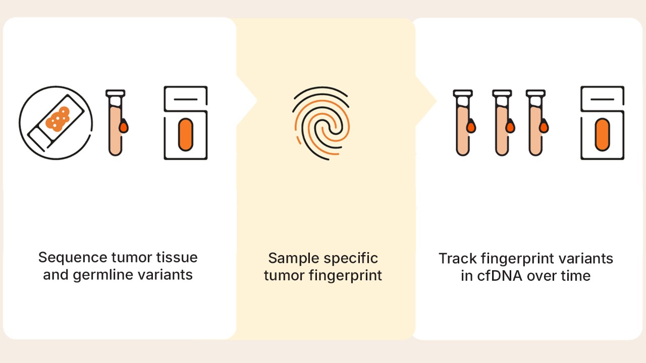
Tumor-informed and tumor-naïve approaches are two different strategies for designing NGS-based assays in MRD research. A tumor-informed strategy begins by profiling a tumor to generate a tumor-specific fingerprint that can be tracked during subsequent MRD analysis. Researchers can use this tumor fingerprint to monitor multiple tumor-specific alterations identified in the original tumor sample.

Tumor-naïve research approaches detect tumor-associated molecular signals directly from plasma without prior tumor sequencing. Without the need for matched tumor sequencing, tumor-naïve methods benefit from a more streamlined detection workflow with variable sensitivity depending on assay design, sequencing depth, and factors affecting ctDNA abundance. As tumors often contain many genomic alterations, monitoring multiple tumor-specific or predefined genomic targets can increase the likelihood of detecting low-abundance ctDNA fragments within the much larger background of non-tumor–derived cfDNA.2,12

How does WGS improve molecular residual disease research?

WGS aggregates signals across thousands of tumor-specific variants. By aggregating these signals, WGS helps overcome sampling limitations and enables ultrasensitive cancer monitoring research. In low disease–burden settings, low tumor fraction and the limited number of cfDNA fragments available for sequencing can constrain detection sensitivity. Additionally, tracking a small number of variants can miss signal when very few tumor-derived fragments are available in a given sample. By monitoring thousands of variants, WGS can increase the likelihood of detecting low-abundance ctDNA.6

WGS tumor-informed approach

Infographic displaying figure 1: Whole-genome sequencing tumor-informed approach workflow.

Figure 1: Whole-genome sequencing tumor-informed approach workflow.

Advantages of WGS for MRD research

By integrating signals across thousands of tumor-specific variants, WGS provides several advantages for tumor-informed MRD research.

Genome-wide variant coverage

Tracks thousands of tumor-specific variants rather than a limited set of predefined targets

Improved sensitivity at low ctDNA levels

Aggregates signal across many variants, increasing the likelihood of detecting rare ctDNA fragments

Multiple genomic signal types

Interrogates multiple classes of genomic alterations within a single sequencing strategy

Flexible analytical framework

Supports integration of additional molecular signals and analytical approaches

Lab tech MRD research

For laboratories developing MRD research capabilities, the decision is not only about sensitivity, but also implementation, scalability, data ownership, and long-term flexibility. As the leading provider of high-throughput sequencing technologies with over 2000 NovaSeq Systems across the globe, Illumina empowers local laboratories to adopt sequencing-intensive applications such as WGS MRD research with greater speed and accessibility.

Illumina molecular residual disease research portfolio

Illumina offers complementary WGS workflows for highly sensitive solid tumor MRD research, including standard sequencing workflows with performance down to 10 parts per million (ppm) and duplex sequencing workflows capable of single-digit ppm detection. Beyond sequencing, MRD research programs require an integrated approach. Illumina delivers a fully connected platform to support performance, scalability, and evidence generation.

Explore the MRD research portfolio

Learn more about Illumina workflow modalities for WGS-based MRD research and explore early access opportunities.* Submit the form to contact our team and receive updates on program availability.

*Early access availability may be limited and subject to program criteria.

MRD research portfolio

MRD FAQ

There is no universally recommended sequencing depth that is appropriate for all molecular residual disease research applications. Sequencing depth requirements depend on the research application and assay design. Important factors include the amount of input DNA, the abundance of tumor-derived molecules, and the desired analytical sensitivity. Greater sequencing depth can increase the opportunity to detect low-abundance ctDNA within the background of non-tumor–derived cfDNA, provided that sufficient unique DNA molecules are available. Additional considerations that influence detection sensitivity include library complexity, background error rates, and error-suppression approaches such as molecular barcoding and duplex sequencing.2,13,14

Numerous factors influence the analytical sensitivity of NGS-based molecular residual disease detection. These factors include the amount of tumor-derived DNA in the sample, the number of unique DNA molecules interrogated by the assay, assay design, and sequencing depth. The sensitivity of NGS-based MRD detection is also influenced by error correction and technical error rates, as errors can interfere with detecting variants present at low frequencies.2


Learn how NGS and DRAGEN analysis reduce noise and errors to enable accurate somatic variant calling in ctDNA.

There is no universal number of tumor mutations that should be tracked for molecular residual disease research. The appropriate number of tumor mutations to track depends on assay design, sequencing approach, and the number of tumor-specific variants available for a given tumor. Researchers can increase the likelihood of detecting ctDNA by tracking numerous tumor-derived variants, especially when ctDNA is present at very low abundance.2,12


Watch the DRAGEN secondary analysis webinar to learn about easy-to-use oncology applications for WGS and a new analysis pipeline for MRD, enabling simplified and streamlined oncology workflows. 

Large-scale MRD research requires sequencing platforms that can generate the data volumes needed to support high-throughput sequencing workflows. Production-scale sequencing systems, such as the NovaSeq X Sequencing System, can support whole-genome sequencing at the throughput required for large-scale studies. WGS approaches can interrogate the genome broadly, enabling MRD analysis methods to aggregate signals across many tumor-derived variants and helping research labs scale tumor-informed WGS MRD workflows.6

Join Dr Alvaro Hernandez from the University of Illinois Urbana-Champaign as he provides insight for scaling NGS in core labs and shares run performance data in this webinar featuring the NovaSeq X Series.


Explore Illumina sequencing platforms for benchtop and production-scale sequencing system specifications to help identify the best sequencer that matches your NGS requirements.

No. NGS-based MRD approaches differ in how they track tumor-specific variants in individual tumor samples. Personalized panel approaches, sometimes described as bespoke, design and manufacture a custom assay based on the molecular profile of each tumor. This can enable highly specific tumor-informed tracking but adds a per-tumor design-and-build step that can increase turnaround time and laboratory operational complexity.

Other tumor-informed approaches use standardized workflows that do not require manufacturing a new panel for each tumor sample. Whole-genome sequencing (WGS), for example, can provide a tumor-informed, panel-free approach that uses a consistent library prep to aggregate signal across thousands of tumor-specific variants across the genome.15

/ Results

Related resources

Demystifying MRD research

Learn from a panel of experts as they discuss WGS-based MRD approaches, best practices, and technical considerations.

WGS solution for MRD research

Discover how Illumina is introducing the first distributed whole-genome sequencing solution for highly sensitive MRD research.

DRAGEN bioinformatics

Learn about expanded research support for MRD and oncovirus workflows and various other DRAGEN capability advancements.

Cancer whole-genome sequencing

Explore library prep, sequencing, and data analysis research options for cancer whole-genome sequencing and tumor–normal comparisons.

Speak with a specialist

Talk to an expert to learn more about our MRD research portfolio.

Early access availability may be limited and subject to program criteria.

References

  1. Jantus-Lewintre E, Calabuig-Fariñas S, Gandara D, et al. Harmonizing definitions in liquid biopsy: A terminology framework by the international society of liquid biopsy. J Liq Biopsy. 2026;12:100462. Published 2026 Mar 25. doi:10.1016/j.jlb.2026.100462
  2. Moding EJ, Nabet BY, Alizadeh AA, Diehn M. Detecting Liquid Remnants of Solid Tumors: Circulating Tumor DNA Minimal Residual Disease. Cancer Discov. 2021;11(12):2968-2986. doi:10.1158/2159-8290.CD-21-0634
  3. Wan JCM, Massie C, Garcia-Corbacho J, et al. Liquid biopsies come of age: towards implementation of circulating tumour DNA. Nat Rev Cancer. 2017;17(4):223-238. doi:10.1038/nrc.2017.7
  4. Pantel K, Alix-Panabières C. Minimal residual disease as a target for liquid biopsy in patients with solid tumours. Nat Rev Clin Oncol. 2025;22(1):65-77. doi:10.1038/s41571-024-00967-y
  5. Baden J, Lin CJ, Anfora AT, et al. Generic Protocols for Analytical Validation of Tumor-Informed Circulating Tumor DNA Assays for Molecular Residual Disease: The Blood Profiling Atlas in Cancer's Molecular Residual Disease Analytical Validation Working Group Consensus Recommendation. JCO Precis Oncol. 2026;10:e2500267. doi:10.1200/PO-25-00267
  6. Zviran A, Schulman RC, Shah M, et al. Genome-wide cell-free DNA mutational integration enables ultra-sensitive cancer monitoring. Nat Med. 2020;26(7):1114-1124. doi:10.1038/s41591-020-0915-3
  7. Widman AJ, Shah M, Frydendahl A, et al. Ultrasensitive plasma-based monitoring of tumor burden using machine-learning-guided signal enrichment. Nat Med. 2024;30(6):1655-1666. doi:10.1038/s41591-024-03040-4
  8. Chen K, Yang F, Shen H, et al. Individualized tumor-informed circulating tumor DNA analysis for postoperative monitoring of non-small cell lung cancer. Cancer Cell. 2023;41(10):1749-1762.e6. doi:10.1016/j.ccell.2023.08.010
  9. Santonja A, Cooper WN, Eldridge MD, et al. Comparison of tumor-informed and tumor-naïve sequencing assays for ctDNA detection in breast cancer. EMBO Mol Med. 2023;15(6):e16505. doi:10.15252/emmm.202216505
  10. Zhang JT, Lin BX, Zhao XG, Zhong WZ, Wu YL. Circulating tumor DNA analysis in lung cancer: from molecular origins to analytical boundaries. Lung Cancer. 2026;217:109473. doi:10.1016/j.lungcan.2026.109473
  11. de Abreu AR, Wyninckx A, Vandamme T, et al. Circulating Tumor DNA detection in cancer: a comprehensive overview of current detection methods and prospects. Oncologist. 2025;30(9):oyaf204. doi:10.1093/oncolo/oyaf204
  12. Li X, Liu T, Bacchiocchi A, et al. Ultra-sensitive molecular residual disease detection through whole genome sequencing with single-read error correction. EMBO Mol Med. 2024;16(9):2188-2209. doi:10.1038/s44321-024-00115-0
  13. Gydush G, Nguyen E, Bae JH, et al. Massively parallel enrichment of low-frequency alleles enables duplex sequencing at low depth. Nat Biomed Eng. 2022;6(3):257-266. doi:10.1038/s41551-022-00855-9
  14. McNulty SN, Mann PR, Robinson JA, Duncavage EJ, Pfeifer JD. Impact of Reducing DNA Input on Next-Generation Sequencing Library Complexity and Variant Detection. J Mol Diagn. 2020;22(5):720-727. doi:10.1016/j.jmoldx.2020.02.003
  15. Frydendahl A, Nors J, Rasmussen MH, et al. Detection of circulating tumor DNA by tumor-informed whole-genome sequencing enables prediction of recurrence in stage III colorectal cancer patients. Eur J Cancer. 2024;211:114314. doi:10.1016/j.ejca.2024.114314