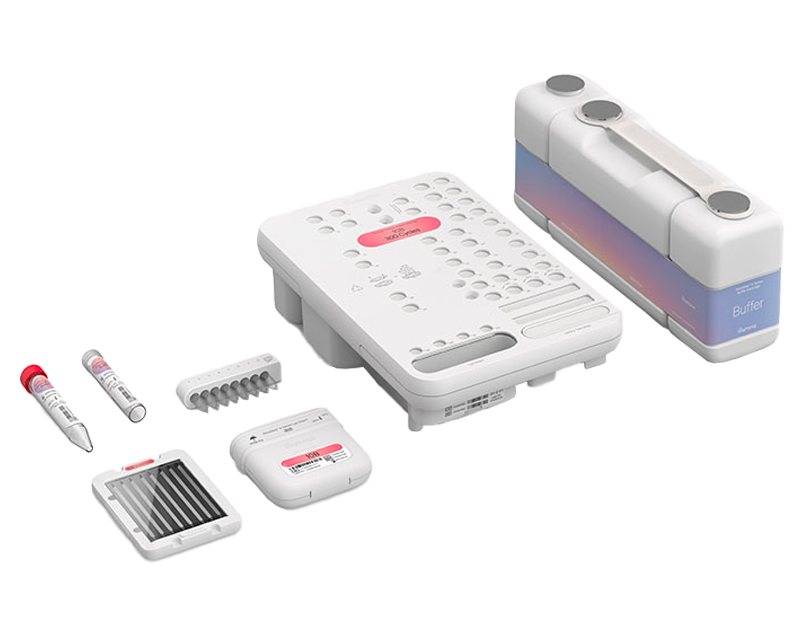
NovaSeq X Series Reagent Kits
Reagent kits for the NovaSeq X Series include a reagent cartridge, buffer cartridge, flow cell, lyo insert, pre-load buffer, and library tube strip.
StrataMap Spatial delivers unbiased whole‑transcriptome analysis with cellular resolution directly within intact tissue.
Assay time
Hands-on time
Input quantity
StrataMap Spatial enables high-resolution, spatially resolved whole transcriptome analysis within intact fresh frozen tissue sections.
Unbiased whole‑transcriptome profiling
Capture gene expression across intact tissue without predefined gene panels, enabling unbiased, spatial discovery.
High sensitivity for rare cell detection
Detect distinct or rare cell populations through improved transcript recovery and whole transcriptome analysis
Versatile project design
Scale easily from pilot projects to large cohort studies by running single slides or batches within one experiment. The 15 mm × 50 mm capture area supports profiling one continuous tissue section or multiple discrete tissue sections.
| Analytical sensitivity | Sequencing-based spatial transcriptomics using poly(A)-capture chemistry |
|---|---|
| Assay time | 2.5 days |
| Content specifications | Coding and long, non-coding RNA with poly(A) |
| Description | StrataMap Spatial enables whole-transcriptome spatial profiling within intact tissue, combining broad gene expression analysis with spatial context to support discovery-driven research, tissue characterization, and multiomic analysis workflows. |
| Hands-on time | ~ 8.5 hr |
| Input quantity | 1-12 sections |
| Instruments | NextSeq 2000 System, NovaSeq X System, NovaSeq 6000 System, NovaSeq X Plus System |
| Method | Whole-transcriptome sequencing |
| Multiplexing | Multiplexing by each slide, not cross slides |
| Nucleic acid type | RNA |
| Sample type details | StrataMap Spatial is compatible with fresh-frozen tissues embedded in optimal cutting temperature (OCT) compound. |
| Specialized sample types | Not FFPE-compatible |
| Species category | Other, Mammalian, Mouse, Canine, Yeast, Human, Rat |
| Species details | Eukaryotic |
| Technology | Sequencing |
For all users:
Illumina Imaging Qualification / Rehearsal slide (optional; required for first-time users)
For cloud users:
ICM professional license (trial version available) or
ICM basic license or
ICA basic license
For local users:
V4 DRAGEN server
For users sequencing in lab:
NovaSeq X 10B or 25B 100c kits
NovaSeq 6000 S2 100c or S4 200c its
NextSeq 2000 P4 100c kits
StrataMap Spatial offers a truly unbiased driven whole transcriptome solution for eukaryotic tissues, supporting applications such as tissue atlasing, biomarker discovery, and tissue microenvironment studies across healthy and diseased samples.
StrataMap Spatial
| Instrument | Recommended number of samples | Read length |
|---|---|---|
| NovaSeq X Plus System | # slides per run: 10B: 1 (L) slide or more; 25B 2 L slides or more |
130+8 (optional) |
| NovaSeq 6000 System | # slides per run: S2: 1 (S) slide or more; S4: 1 L slide or more |
130+8 (optional) |
| NextSeq 2000 System | # slides per run: P4 0.5 - 1 (S) slide |
130+8 (optional) |
Next Generation Sequencing (NGS)
Discover the broad range of experiments you can perform with next-generation sequencing, and find out how Illumina NGS works.
Single-Cell and Ultra-Low-Input RNA-Seq
With single-cell RNA-Seq, or scRNA-Seq, you can study cellular differences often masked by bulk sampling. Explore high- and low-throughput single-cell sequencing methods.
Map transcriptional activity within structurally intact tissue to unravel complex biological interactions using spatial RNA-Seq.
A dedicated support section is not currently available for this product
, cancer-associated fibroblasts (red), and pro-tumor macrophages (purple) throughout the tumor tissue is shown.")
StrataMap Spatial reveals cellular diversity
Whole-transcriptome spatial profiling of high-grade invasive ductal carcinoma resolves gene expression across the tumor microenvironment. Expansion of proliferating tumor cells (aqua), cancer-associated fibroblasts (red), and pro-tumor macrophages (purple) throughout the tumor tissue is shown.

StrataMap Spatial enhances differential gene expression detection
Whole-transcriptome spatial profiling vs 5K (top) and 18K (bottom) commonly used gene panels. Of all upregulated biomarkers detected by StrataMap Spatial, (A) 63.6% and 13.0% at P > 0.001, and (B) 63.8% and 14.6% at P > 0.05 were missed by the respective panels.

Cellular-resolution spatial mapping reveals cell populations and localized gene expression
Localized gene expression at cellular resolution in ductal carcinoma in situ tissue. (A) Cell-type visualization shows fibroblasts (yellow) in normal stroma and myofibroblasts (red) at the tumor–stromal interface. (B) Spatial expression of ACTA2, a myofibroblast biomarker, in the same tissue section.

Sensitive transcript detection across tissue states with StrataMap Spatial
Spatial transcriptomic profiling of ductal carcinoma in situ and grades 1–3 invasive ductal carcinoma shows consistent transcript detection across disease states. H&E images align with spatial heatmaps, linking morphology and gene expression.

Large-format spatial capture supports multi-sample tissue profiling
StrataMap Spatial provides a 750 mm² capture area, providing broader coverage. (A) Comparison of spatial capture areas across spatial transcriptomics platforms. (B) Transcriptional changes between virgin and pregnant mouse heart tissue sections.

Illumina Connected Multiomics enables integrated spatial analysis
Analysis outputs generated in Illumina Connected Multiomics from a mouse kidney sample. (A) Spatial domains. (B) Sankey diagram. (C) UMAP plot. (D) Transcript heatmap. (E) Violin plots. (F) Gene expression heatmap.

On-demand webinar of ASHG 2025 presentation

On-demand webinar of AGBT 2026 Gold Sponsor Workshop
StrataMap Spatial utilizes a large, advanced tissue placement area with continuous 1 µm surface features.
Captured transcripts are binned with integrated cell segmentation through the Illumina spatial analysis pipeline.
The StrataMap Spatial assay is optimized for use with fresh-frozen tissue. A version supporting formalin-fixed, paraffin-embedded (FFPE) tissue is in development.
Two slide formats are available: a large-format slide with a 50 × 15 mm capture area and a second format with a 17 × 15 mm capture area. Both use a standard 75 × 25 mm microscope slide.
Coverage varies based on tissue type, tissue quality, and sequencing depth. During product development, StrataMap Spatial detected > 2× more genes than panel-based spatial solutions.
Detection sensitivity can vary by tissue type, development stage, disease type, and sample quality or age. During product development, StrataMap Spatial detected up to 4000 unique transcripts per 10 × 10 µm bin.
Reagent kits for the NovaSeq X Series include a reagent cartridge, buffer cartridge, flow cell, lyo insert, pre-load buffer, and library tube strip.
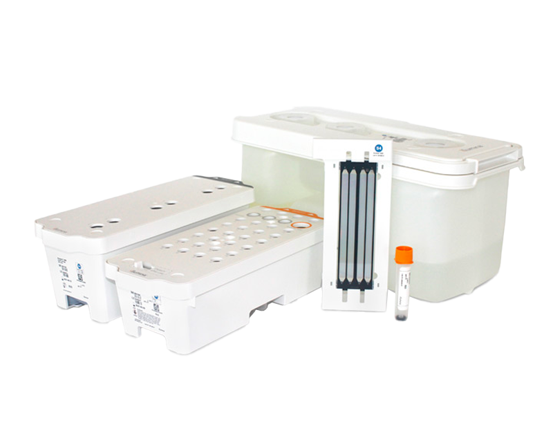
Reagent kits for the NovaSeq 6000 System consisting of ready-to-use pre-filled reagent cartridges and flow cells.
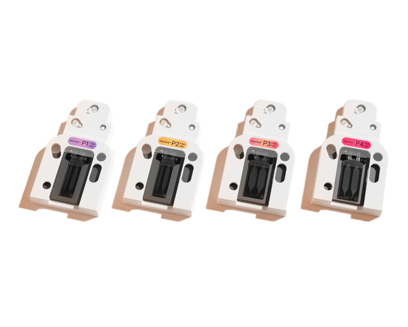
Reagents for the NextSeq 1000/2000 System feature easy-to-use cartridges and multiple flow cell configurations for flexible sequencing options.

Illumina Connected Multiomics is a powerful and intuitive cloud-based solution that enables multiomic data analysis and visualization at scale.

Accessible and highly scalable single-cell RNA-Seq solution for mRNA capture, barcoding, and library prep without complex workflows or microfluidics
A rapid, cost-effective workflow for accurate, unbiased detection of the protein-coding transcriptome with precise measurement of strand information.

A single assay for comprehensive discovery of the 5-base genome (A, T, G, C, and 5mC), providing dual insights into the whole genome and methylome.

Highly accurate, comprehensive germline human whole-genome sequencing with only ~10 min of hands on time, without traditional library prep.
Reach out for information about our products and services, or get answers to questions about our technology.